

Diagnostic Imaging
Elite sport horses demand a standard of care that keeps pace with what’s asked of them, and our diagnostic imaging is often where that care begins. At Metamora Equine, Roland Thaler, VMD DACVSMR brings over 40 years of equine sports medicine experience to every evaluation, including diagnostic imaging that often determines what happens next for your horse. Precise imaging is not just a tool for spotting problems. It’s how informed veterinary decisions get made. When you need answers beyond what a physical exam can provide, diagnostic imaging gives you and your veterinarian the clarity to act with confidence.
How Digital Radiography and Ultrasound Confirm a Diagnosis
When a horse shows signs of lameness, swelling, or a drop in performance, a physical exam can only reveal so much. Digital radiography and ultrasound fill in what hands and eyes cannot detect. Radiographs produce clear images of bone, showing fractures, bone remodeling, joint space narrowing, and calcification. Ultrasound captures soft tissue in real time, making it possible to visualize tendons, ligaments, joint capsules, and bursae in detail that no other field-based tool can match.
Together, these two modalities confirm what a clinical exam suspects. A suspected suspensory injury gets visualized and measured. A subtle bony change in the coffin joint gets documented. Neither tool replaces the other. Radiography and ultrasound answer different questions, and a thorough diagnostic approach uses both where appropriate. The result is a clear, evidence-based picture of what is actually happening inside the horse, which is the only reliable foundation for an effective treatment plan.
Benefits of Diagnostic Imaging
Accurate Identification of Injury Location and Severity
Pinpointing the source of pain or dysfunction is the first step toward effective treatment. Diagnostic imaging removes the guesswork by providing a direct view of bone, joint, and soft tissue structures. This precision means your horse is not subjected to treatments targeting the wrong area, saving time and reducing unnecessary stress on the animal.
Informed Treatment Planning from the Start
When your veterinarian can see exactly what is happening internally, treatment decisions become more targeted and effective. Imaging findings shape everything from medication protocols to rehabilitation plans and activity restrictions. Rather than relying solely on clinical signs, your care team has concrete data to build from, which typically leads to better outcomes and fewer setbacks.
Early Detection of Developing Conditions
Performance horses often mask discomfort until a problem becomes significant. Diagnostic imaging can reveal early-stage changes in bone density, joint surfaces, or tendon integrity before they escalate. Catching these issues early gives you more options and more time, potentially preventing minor concerns from becoming career-ending injuries.
Objective Progress Monitoring During Recovery
Once a treatment plan is underway, imaging allows your veterinarian to assess how well the horse is healing. Rather than estimating recovery based on behavior alone, follow-up scans provide measurable data on tissue repair and structural integrity. This supports smarter return-to-work timelines and reduces the risk of reinjury from returning to work too soon.
Seamless Integration with a Complete Diagnostic Plan
Diagnostic imaging does not exist in a silo. Every scan or radiograph feeds into a comprehensive evaluation that considers the horse’s full history, workload, and goals. This connected approach ensures that imaging findings translate directly into actionable clinical decisions rather than isolated data points sitting in a file.
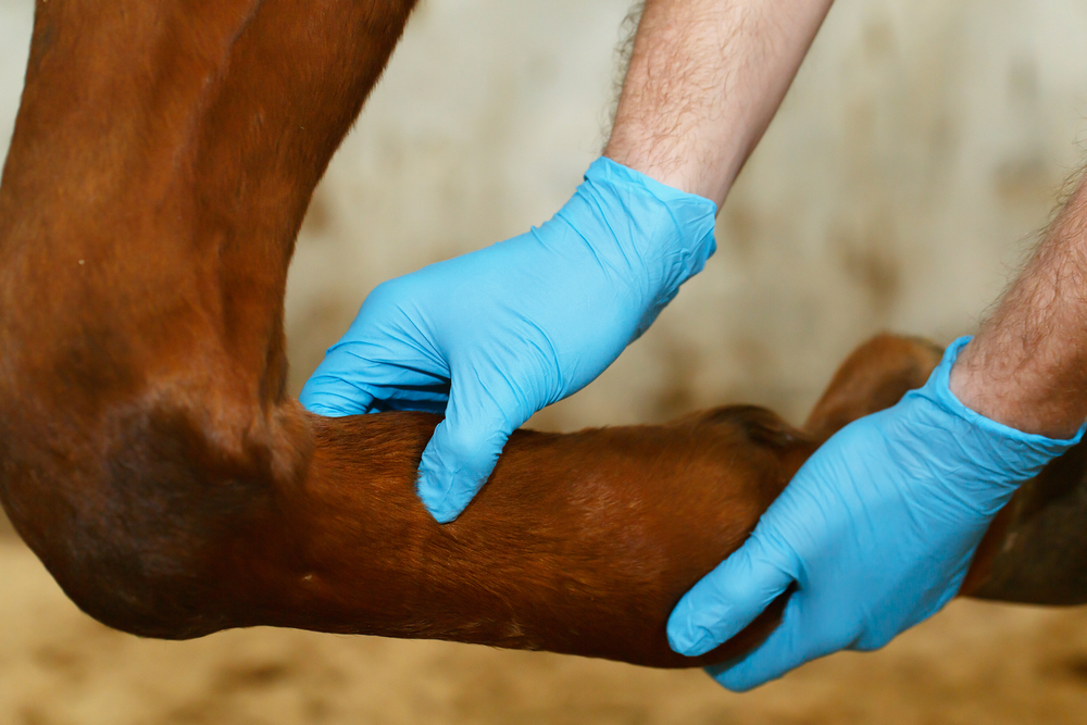
What to Expect During Your Imaging Appointment
Why Choose Us for Diagnostic Imaging
What you get from a diagnostic imaging appointment here is not just a set of images; it is a clear explanation of what those images mean for your horse and what should happen next. Owners and trainers who work with Roland Thaler, VMD DACVSMR leave appointments with specific answers, not possibilities. That level of clarity comes from decades of working exclusively with performance horses across competitive environments in Michigan, Ohio, North Carolina, and South Carolina, where the stakes attached to every diagnosis are high and vague answers are not acceptable.
The credentials Dr. Thaler holds, including Diplomate of the American College of Veterinary Sports Medicine and Rehabilitation and Certified ISELP Member, reflect a standard of clinical knowledge that directly shapes how imaging findings are interpreted and applied. His standing on professional advisory boards and his history as an early adopter of advanced veterinary technology mean your horse’s imaging is read by someone operating at the leading edge of the field. That translates to more accurate diagnoses, better-informed treatment decisions, and a higher level of confidence for you as the owner.

Schedule Our Diagnostic Imaging Evaluation Today
Our diagnostic imaging is one of the most valuable things you can have heading into a competition season. Roland Thaler, VMD DACVSMR, and the team at Metamora Equine are ready to give your horse the precise, imaging-backed evaluation that serious sport horse care demands, so you can move forward with a plan grounded in facts, not guesswork. Your horse’s health and performance are too important to leave to uncertainty.
Schedule your appointment by dialing 248.310.5568; the team is ready to get your horse on the books. For equine health tips, practice updates, and a closer look at the work done for sport horses across the Midwest, find Metamora Equine on Facebook and follow along between visits.
FAQs
How much is an equine ultrasound?
The cost of an equine ultrasound varies depending on the region, the number of structures being evaluated, and whether it is part of a broader lameness examination. In general, equine ultrasound can range from a few hundred dollars for a focused scan to more for a comprehensive multi-structure evaluation. Your veterinarian will outline expected costs during the consultation so there are no surprises.
How to read an equine ultrasound?
Equine ultrasound images display soft tissue structures in varying shades of gray, with denser tissues appearing brighter and fluid-filled areas appearing darker or black. Tendons and ligaments have a characteristic fiber pattern that trained veterinarians use to identify disruptions, tears, or areas of reduced echogenicity indicating damage. Interpreting these images accurately requires significant clinical experience, which is why findings should always be reviewed and explained by a qualified equine veterinarian.
How to ultrasound equine tendons?
Ultrasounding equine tendons requires clipping the hair over the area of interest, applying acoustic coupling gel, and using a high-frequency linear probe to image the tendon in both transverse and longitudinal planes. The veterinarian systematically scans the tendon from top to bottom, looking for changes in fiber alignment, echogenicity, or cross-sectional area that may indicate injury. This process requires precise probe positioning and clinical expertise to ensure nothing is missed and findings are interpreted correctly.
How to prepare for an ultrasound on a horse?
Preparation for an equine ultrasound typically involves clipping the hair from the area to be imaged to improve contact and image quality. The skin should be clean and free of dirt, debris, or wound treatment products that could interfere with the scan. Your veterinarian may also ask you to avoid applying liniments or ointments to the area in the days before the appointment, so check with the clinic beforehand if you have any questions.